Understanding Pasteurellosis (Fowl Cholera) in Poultry: Causes, Symptoms, and Management
Pasteurellosis, also known as Fowl Cholera, is an acute septicemic disease affecting domestic fowl and wild birds caused by Pasteurella multocida. This disease is characterized by high morbidity and mortality rates, primarily impacting chickens, turkeys, and ducks. The disease spreads through healthy carriers, ingestion, inhalation, and mechanical transmission. Symptoms vary from sudden death in well-fleshed birds to chronic manifestations like swollen wattles, joint inflammation, and torticollis. Detailed examination reveals characteristics such as mucous in the mouth and nostrils, cyanotic comb and wattles, and various internal organ abnormalities. Effective management strategies are essential to control the spread of Pasteurellosis in poultry populations.

Download Presentation

Please find below an Image/Link to download the presentation.
The content on the website is provided AS IS for your information and personal use only. It may not be sold, licensed, or shared on other websites without obtaining consent from the author. Download presentation by click this link. If you encounter any issues during the download, it is possible that the publisher has removed the file from their server.
E N D
Presentation Transcript
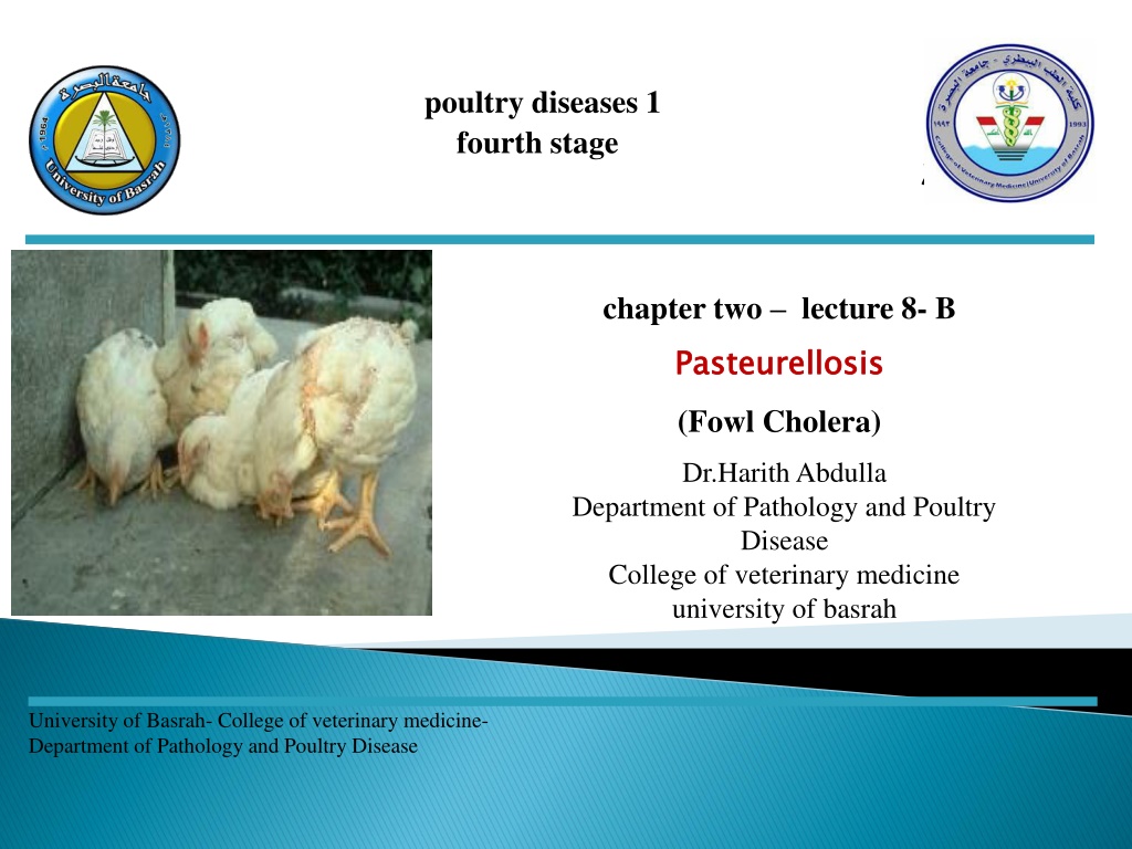
poultry diseases 1 fourth stage chapter two lecture 8- B Pasteurellosis (Fowl Cholera) Pasteurellosis Dr.Harith Abdulla Department of Pathology and Poultry Disease College of veterinary medicine university of basrah University of Basrah- College of veterinary medicine- Department of Pathology and Poultry Disease
(Fowl Cholera) (Fowl Cholera)
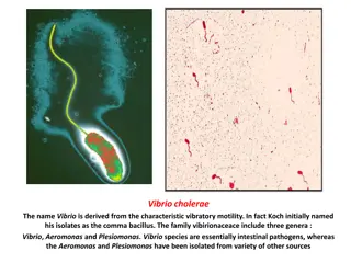
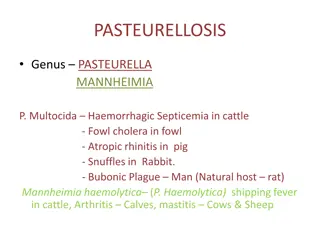
1-Avian Cholera. 2-Avian Pasteurellosis. 3-Avian Hemorrhagic Septicemia. Definition: Fowl cholera is acute septicemic disease of domestic fowl and wild birds caused by Pasteurella multocida, (Pasteurella aviseptica) characterized by high morbidity and mortality. Definition:
Chickens, turkeys and ducks are most commonly affected. Epizootiology: 1-Healthy nasal carriers provide a source of infection. 2-The natural spread of the disease is by ingestion and inhalation. 3-Mechanical transmission by vectors. Epizootiology:
1- Age: Semimature to mature. 2- Acute: a-Sudden death of well fleshed birds. b-Greenish and yellowish diarrhea. c- Mucous in mouth and nostrils. d- Cyanotic comb and wattles. 3-Chronic: a-Carriers due to localization of the bacteria. b-Swollen wattles and eyes. c- Inflammation of joints and tendon sheath of legs and wings. d- Torticollis.
Acute: a- A sticky mucous in the mouth and nasal passages. b- Generalized congestion. C- Hemorrhage in the heart muscles particularly around the coronary groove and gizzard. d- Pericardial sac contains an excess of yellowish fluid . e- Liver: very dark or lighter than usual, with many white necrotic foci. f-Inflammation and hemorrhage in the duodenum. g- Lung: Consolidation and congestion with small hemorrhage . h- Cheesy , yellowish deposits in various parts of the body, especially on the air sacs and intestine.
Chronic type: a-Dried cheesy, yellow material is found free in the abdominal cavity or adhere to some organ due to ruptured yolk. Chronic type: b-Hemorrhage of the ovary . Ova: Soft, flabby, irregular in outline and pedunculated . Greenish colored ovum is observed.(Due to salpingitis). C- Caseous swollen wattles and joints. d- Suppurative meningitis due to the localization of bacteria at the base of skull, ear and brain.
1-Coagulation necrosis. 2-Heterophilic infiltration. Diagnosis: 1-History. 2-Signs. 3-Lesions. 4-Laboratory diagnosis. a-Finding of bipolar bacteria. b- Isolation and characterization of bacteria from circulatory blood, liver and other organs. c- Agglutination test. d- Laboratory animal inoculation ( chick and mice). Diagnosis:
1-Water sanitation. 2- Good management. 3-Control flies and rodents. 4-Vaccination at 12-16 weeks and repeat 4-8 weeks later. Treatment: 1-Sulfanomides. 2-Teramycin in water or feed. Treatment:
Pullorum Pullorum Fowl Typhoid Fowl Typhoid Fowl Fowl Cholera Cholera Comb and wattles Pale Pale Cyanotic Joints Normal Normal Swollen Liver size Enlarged 2-3 Enlarged 2-3 Normal to slightly enlarged Red Liver color Red Bronzy Necrotic foci in the liver Spleen size + + ++ Enlarged 2-3 Enlarged 2-3 Normal Heart Nodules Nodules Hemorrhagic Ceca Core Core Inflammation and hemorrhage

")