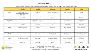
Select Reps for Feedback Lunch

This collection features detailed images of various types of epithelium found in different parts of the body, including the bladder, submandibular gland, ileum, uterine tube, esophagus, skin, foot, and trachea. Each image provides a close-up view of the epithelial tissue and surrounding structures, highlighting the unique characteristics and functions of each type. These slides serve as valuable educational resources for studying histology and understanding the histological makeup of different organs.

Download Presentation

Please find below an Image/Link to download the presentation.
The content on the website is provided AS IS for your information and personal use only. It may not be sold, licensed, or shared on other websites without obtaining consent from the author. Download presentation by click this link. If you encounter any issues during the download, it is possible that the publisher has removed the file from their server.
E N D
Presentation Transcript
WebSlide 0098_G: Bladder (simple sq. epithelium, transitional epithelium) serosa SiSE SiSE lumen DICT TrE LCT DICT TrEc
WebSlide 0038_G: Submandibular gland (simple cuboidal epithelium lining secretory ducts) ducts in longitudinal section ducts in cross section
WebSlide 0032_G: Ileum (simple columnar epithelium) serosa lumen LCT tb s(b)b epith gc gc gc LCT epith LCT
WebSlide 0051_G: Uterine Tube (simple columnar ciliated epithelium) lumen sm basal bodies cilia ncc epith
WebSlide 0078A_G: esophagus (stratified sq. non-keratinized epithelium) basal cells surface squamous cells
WebSlide 0065_G: skin, foot (stratified sq. keratinized epithelium) sweat gland duct look along this boundary to see sweat glands & ducts basal cell layer (note ABSENCE of nuclei in the stratum lucidum and corneum)
WebSlide 0008_G: trachea (pseudostratified ciliated columnar epithelium) tracheal gland secretory duct basal cells basement membrane ciliated columnar epithelial cell (note height of cilia) goblet cell (note ABSENCE of cilia)
Slide UMich #30: abdominal mesentery examples of simple squamous epithelium: mesothelium endothelium
Slide UMich 9N-1: Kidney (simple cuboidal epithelium, coiled tubes in various planes of section)
Slide UMich 9N-1: Kidney (simple cuboidal epithelium, tubes mostly in longitudinal section)
Slide UMich 153: Esophagus (stratified squamous non-keratinized epithelium) GL(sce) adventitia LCT lumen duct duct DICT artificial separation LCT SM DICT StSE lumen StSEc
Slide UMich 106: Thick Skin (stratified squamous keratinized epithelium) Deep fascia Deep fascia connective tissue (dermis) blood vessel epithelium (epidermis) keratinized layer
Slide UMich 40: trachea (pseudostratified columnar ciliated epithelium with goblet cells) Cilia Basal bodies Goblet cell Basement membrane


")


")