Pediatric Emergency Management Guidelines
A case study of an 11-month-old boy presenting with cough, fever, and respiratory distress, highlighting the stages in the management of a sick child including triage, emergency treatment, history/examination, laboratory investigations, diagnosis, treatment, and more. It emphasizes the importance of quickly assessing for emergency signs, identifying priority and emergency signs in pediatric patients, and providing oxygen therapy effectively.
Download Presentation

Please find below an Image/Link to download the presentation.
The content on the website is provided AS IS for your information and personal use only. It may not be sold, licensed, or shared on other websites without obtaining consent from the author. Download presentation by click this link. If you encounter any issues during the download, it is possible that the publisher has removed the file from their server.
E N D
Presentation Transcript
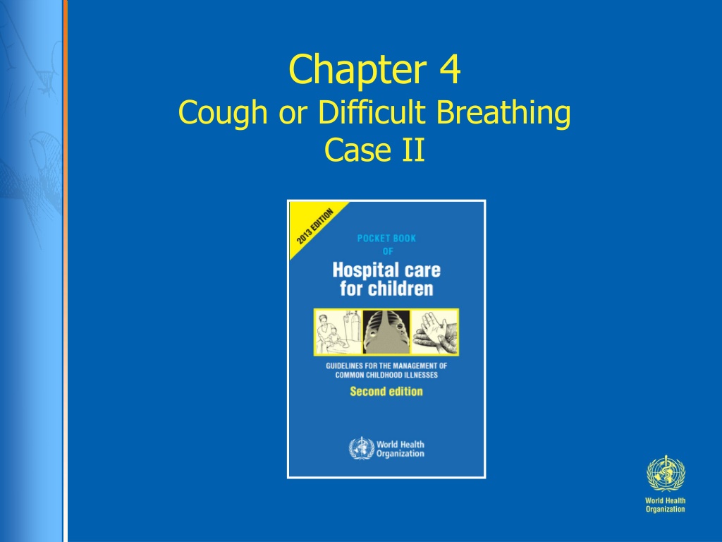
Chapter 4 Cough or Difficult Breathing Case II
Ratu 11 month old boy with 5 days of cough and fever, yesterday he became short of breath and unable to feed
What are the stages in the management of any sick child?
Stages in the management of a sick child (Ref. Chart 1, p. xxii) 1. Triage 2. Emergency treatment 3. History and examination 4. Laboratory investigations, if required 5. Main diagnosis and other diagnoses 6. Treatment 7. Supportive care 8. Monitoring 9. Discharge planning 10. Follow-up
At Triage how to quickly assess for emergency signs Take a brief history of the presenting problem Take temperature and weigh the child A. Listen for stridor or obstructed breathing B. Look for cyanosis and for signs of respiratory distress (chest indrawing, tracheal tug), check SpO2 C. Feel the skin temperature of the hands and feet, feel the peripheral pulses for volume, check capillary refill time D. Assess for lethargy and level of interaction.
Do you notice any emergency or priority signs? Temperature: 39.70C, pulse: 180/min, RR: 70/min, cyanosis visible suprasternal and subcostal recession, grunting respiration
Triage Priority signs (Ref. p. 6) Tiny baby Temperature Trauma Pallor Poisoning Pain (severe) Respiratory distress Restless, irritable, lethargic Referral Malnutrition Oedema of both feet Burns Emergency signs (Ref. p. 2, 6) Obstructed breathing Severe respiratory distress Central cyanosis Signs of shock Coma Convulsions Severe dehydration
Triage Priority signs (Ref. p. 6) Tiny baby Temperature Trauma Pallor Poisoning Pain (severe) Respiratory distress Restless, irritable, lethargic Referral Malnutrition Oedema of both feet Burns Emergency signs (Ref. p. 2, 6) Obstructed breathing Severe respiratory distress Central cyanosis Signs of shock Coma Convulsions Severe dehydration
How to give oxygen Use an 8 F size tube Measure the distance from the side of the nostril to the inner eyebrow margin with the catheter Insert the catheter to this depth and secure it with tape Place the prongs just inside the nostrils and secure with tape. (Ref. Chart 5, p. 11 p. 312-315) Start oxygen flow at 1-2 litres/minute, in young infants at 0.5 litre/minute
Emergency treatment (continued) Blood glucose 1.8 mmol/l: How do you treat hypoglycaemia? Give IV glucose (Ref. Chart 10, p. 16)
History Ratu is a 11 month old boy with 5 days of cough and fever. Yesterday he became short of breath and was unable to feed. He was well until 5 days ago. Then he developed fever with cough. He was taken to a local medical shop, where he was given two types of syrupy medicine. He deteriorated over two days with high fever, increased difficulties in breathing and today he cannot feed. No significant past illnesses Family history: Ratu's grandmother had tuberculosis, which was treated 3 years ago. Social history: he lives with his parents and grandmother in a small semi-permanent house
Examination Ratu was pale, ill-looking and cyanosed. He had fast breathing with visible suprasternal and subcostal recession and with grunting respiration. Vital signs: temperature: 39.70C, pulse: 180/min, RR: 70/min Oxygen saturation SpO2 : 93% on oxygen Weight: 8.2 kg (check z-score, p 384) Chest: bilateral course crepitations with suprasternal and subcostal recession, grunting and wheeze Cardiovascular: capillary refill 2 seconds, three heart sounds were heard with gallop rhythm; the apex beat was displaced to the left anterior axillary line Abdomen: liver was palpable 4 cm below the right costal margin Neurology: tired but alert; no neck stiffness
Differential diagnoses List possible causes of the illness, in order they are likely, use clinical features to say which are most and least likely (Ref. p. 77-79, p. 93)
Differential diagnoses What clinical features make these diagnoses most or less likely? Pneumonia Malaria Severe anaemia Cardiac failure Congenital heart disease Tuberculosis Pertussis Foreign body Effusion/empyema Pneumothorax Pneumocystis pneumonia (Ref. p. 93, 77-79)
Additional questions on history Immunization history Nutritional history Tuberculosis in family
Additional questions on history Immunization history Nutritional history Breast fed for 3 months, now on powdered cows milk, 2 meals a day, eats fruits (banana, papaya), rarely eats meat or vegetables, some cereals and biscuits
Examination based on possible diagnoses Assess cause of respiratory distress: - Pneumonia: crepitations, bronchial breathing, effusion, cyanosis - Heart failure: tachycardia > 160/min (Ref. p. 120), gallop rhythm, enlarged liver, fast breathing Assess signs and cause of anaemia -Palmer pallor (Ref. p. 121, 199, 307) -If from a malaria area, Look for signs of malaria - Fever, enlarged spleen, anaemia (Ref. p. 156-165) Assess nutritional state - Weight-for-age 8.2kg between -1 and -2 z-scores - Look for oedema of feet (Ref. p. 198)
Further examination based on differential diagnoses Palmar Pallor indicating severe anaemia (Ref. p. 166). Check also conjunctiva and mucous membranes In any child with palmar pallor, check the haemoglobin level
What investigations would you like to do to make your diagnosis?
Investigations Full Blood Examination and blood film Group and cross-match Malaria RDT, thick and thin blood film What are the indications for chest x-ray? Suspicion of effusion, empyema, pneumothorax Unilateral changes on examination Clinical signs of heart failure If tuberculosis is suspected (Ref. p. 77, p. 85) Cardiac echo to look for congenital heart disease
Full blood examination Haemoglobin Platelets WCC Neutrophils Lymphocytes 5.9 g/dl (105-135) 858 x 109/l (150-400) 30.6 x 10⁹/l (6.0-18.0) 26.0 x 10⁹/l (1.0-8.5) 3.4 x 10⁹/l (4.0-10.0)
Blood film: hypochromic microcytic anaemia Hb 5.9g / dL, MCV 62 No malaria parasites, RDT negative
Clinical summary Fever; severe respiratory distress, cyanosis, palmar pallor, bilateral course crepitations, grunting and wheeze; three heart sounds, gallop rhythm and tachycardia Chest x-ray shows enlarged heart and bilateral opacities SpO2 : 82% on room air, 93% on oxygen Hypoglycaemia (1.8 mmol/L, 4.5 mmol/L after glucose) Blood examination shows anaemia (Hb 5.9), neutrophilia, thrombocytosis
Diagnosis Severe pneumonia Heart failure Severe anaemia Severe iron deficiency
Treatment Severe pneumonia Oxygen therapy Antibiotics (Ref. p. 82) (Ref. p. 82) Heart failure Diuretics Fluid restriction (Ref. p. 120-122) Anaemia (with heart failure) Blood transfusion Iron therapy (when improved) Diet change (Ref. p. 307-308)
What supportive care and monitoring are required?
Supportive care Fever management (Ref. p. 305) Fluid management Avoid overhydration: Ratu has very severe pneumonia, heart failure, severe anaemia, and he receives IV therapy and blood transfusion What type of fluid? Nutrition (Ref. p. 294-303) Insert a nasogastric tube and give some feeds What volume of feeds?
Monitoring Use a Paediatric monitoring and response chart to record (Ref. p. 320, 413) Vital signs, respiratory distress, SpO2, AVPU Feeding / nutrition Blood glucose Treatments given Frequency of monitoring: Hourly until out of the red zone 2 hourly until out of orange zone Check by doctor at least 3x per day
Discharge planning and Follow up When is it OK for Ratu to be discharged? What follow-up is needed?
Discharge planning and Follow up When is it OK for Ratu to be discharged? Respiratory distress resolved No hypoxaemia Completed course of parenteral antibiotics Able to take oral medications Check Hb shows improvement Started on iron Cardiac echo normal Parents understand the problems What follow-up is needed Anaemia Nutritional
Summary Seriously ill children may present with one symptom but may have multiple problems: Severe respiratory distress due to: Pneumonia Anaemia, due to iron deficiency Heart failure due to anaemia and severe pneumonia Emergency treatment is life saving Need to identify and treat each problem if the child is to survive Monitoring and supportive care are vital Don t forget follow-up
