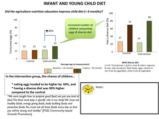
Understanding Erythropoiesis, Iron Metabolism, and Hemoglobin Regulation
Explore the intricate control of erythropoiesis, iron metabolism, and hemoglobin synthesis in the body. Delve into the role of erythropoietin, factors influencing erythropoiesis rate, essential nutrients like iron and vitamins, and clinical conditions associated with anomalies in these processes. Gain insights into anemia, its classification, assessment, and physiological consequences, along with an overview of polycythemia. Enhance your understanding of blood cell production and the vital components involved in maintaining healthy blood function.

Download Presentation

Please find below an Image/Link to download the presentation.
The content on the website is provided AS IS for your information and personal use only. It may not be sold, licensed, or shared on other websites without obtaining consent from the author. Download presentation by click this link. If you encounter any issues during the download, it is possible that the publisher has removed the file from their server.
E N D
Presentation Transcript
Control of Erythropoiesis, Iron Metabolism and Hemoglobin
Objectives; Intended learning outcomes (ILOs) After reviewing the PowerPoint presentation and the associated learning resources, the student should be able to: Discuss regulation of erythropoiesis Describe the factors required for normal erythropoiesis and the role of erythropoietin in erythropoiesis Recognize clinical conditions associated with high level of erythropoietin in the blood Summarize the synthesis of Hemoglobin Recognize hemoglobin structure and its functions Describe metabolism of iron and vitamin B12 Define anemia Classify anemia and explain its assessment Describe the physiological consequences and clinical picture of anemia Define polycythemia Classify polycythemia Describe the physiological consequences of polycythemia
Learning Resources Guyton and Hall, Textbook of Medical Physiology; 13th Edition; Unit VI-Chapter 33.
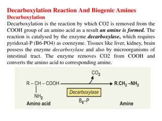
Regulation of Erythropoiesis Erythropoiesis is under the influence of erythropoietin Other Factors Influencing Rate of Erythropoiesis: Hormones: androgens (testosterone), thyroxin, growth hormone and cortisol stimulate erythropoiesis. Deficiency of any of these hormones results in anemia Adequate diet: Amino acids: are important for the formation of globin in Hb. Severe protein deficiency anemia Iron (Fe): 60-70% of iron in the body is in the Hb inside the RBCs. Fe is important for the formation of heme in Hb. Iron deficiency iron-deficiency anemia (microcytic hypochromic anemia) Vitamins: Vitamins B12 and folic acid are important for the synthesis of nucleoproteins (DNA) and final maturation of RBCs. Deficiency of Vitamins B12, folic acid megaloblastic anemia. Pernicious anemia is due to deficiency of Vit. B12 due to lack of intrinsic factor produced by gastric mucosa, which is needed for Vit B12 absorption. Pernicious anemia is an autoimmune condition in which the body's immune system attacks the actual intrinsic factor protein or the parietal cells in the lining of the stomach that make it. Other vitamins :Vit B6, Riboflavin, nicotinic acid, biotin (B7),Vit C, Vit E
Erythropoietin Erythropoiesis is stimulated by erythropoietin Erythropoietin is a glycoprotein, which is synthesized from renal cortex (90%) and the liver (10%) Erythropoietin stimulate the production of proerythroblasts from hematopoietic stem cells in the bone marrow. Once the proerythroblasts are formed, erythropoietin causes these cells to pass more rapidly through the different erythroblastic stages and can increase it to perhaps 10 or more times normal Erythropoietin can be measured in plasma & urine High level of erythropoietin are produced in response to hypoxia ( O2), which can be caused by: Low RBC count (Anemia) Hemorrhage High altitude Prolong heart failure Lung disease
Vitamin B12 and Folic Acid Important for DNA synthesis and final maturation of RBCs. Dietary sources include: meat, milk, liver, fat, green vegetables. Deficiency leads to: Failure of nuclear maturation & division Abnormally large & oval shaped RBCs Short life span of RBCs Reduced RBC count & Hb concentration Macrocytic (megaloblastic) anemia Note the hypersegmented neurotrophiland also that the RBC are almost as large as the lymphocyte. Finally, note that there are fewer RBCs.
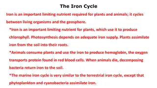
Iron Metabolism Iron is needed for the synthesis of hemoglobin, myoglobin, cytochrome oxidase, peroxidase & catalase The total quantity of iron in the body averages 4-5 g Hemoglobin: Stored iron: Myoglobin: Heme compounds that promote intracellular oxidation (cytochrome): Plasma iron: (transferrin): 65-75% (3 g) 15-30%, particularly in the form of ferritin in the liver, spleen and bone marrow. 4% 1% 0.1% A man excretes about 0.6 mg of iron each day, mainly into the feces. Additional quantities of iron are lost when bleeding occurs. For a woman, additional menstrual loss of blood brings long-term iron loss to an average of about 1.3 mg/day. Diet provides 10-20 mg iron per day. Only 10% of dietary iron is absorbed (1 2 mg ). Iron in food mostly in oxidized form (Ferric, F+3). It is better absorbed in reduced form (Ferrous, F+2). Iron in stomach is reduced by gastric acid, and Vitamin C.
Iron is absorbed from all parts of the small intestine, Iron Absorption, Transport and Metabolism The liver secretes apotransferrin into the bile. In the duodenum, the apotransferrin binds with iron forming transferrin. The formed transferrin is absorbed into in the intestinal epithelial cells by receptor- mediated pinocytosis. This is later released into the blood capillaries in the form of plasma transferrin. Only a few milligrams of iron can be absorbed per day. Thus, even when tremendous quantities of iron are present in the food, only small proportions can be absorbed.
Iron Absorption, Transport and Metabolism When iron is absorbed from the small intestine, it immediately combines in the blood plasma with apotransferrin, to form transferrin, which is then transported in the plasma. The iron is loosely bound in the transferrin and, consequently, can be released to any tissue cell at any point in the body. Excess iron in the blood is deposited and stored especially in the liver hepatocytes and less in the Reticuloendothelial cells of the bone marrow. In the cell cytoplasm, iron combines mainly with a protein, apoferritin, to form ferritin. This iron stored as ferritin is called storage iron. Smaller quantities of the iron in the storage pool are in an extremely insoluble form called hemosiderin. This is especially true when the total quantity of iron in the body is more than the apoferritin storage pool can accommodate.
Iron Absorption, Transport and Metabolism When the body has become saturated with iron so that all apoferritin is already combined with iron, the rate of additional iron absorption from the intestinal tract becomes greatly decreased. Conversely, when the iron stores have become depleted, the rate of absorption can accelerate probably five or more times normal. Thus, total body iron is regulated mainly by altering the rate of absorption.
Serum Iron and Total Iron Binding Capacity (TIBC) Normally transferrin in the plasma is 30-40% saturated with iron, giving a plasma iron level of 100-130 g/100ml) When transferrin is 100% saturated with iron plasma iron level reaches 300 g/100ml. This is referred to as Total Iron Binding Capacity (TIBC)
Anemia and Polycythemia Anemia is decrease in RBC mass as determined by Hct or Hb values below reference level for age and gender. The major causes of anaemia are: 1. RBC Loss without RBC destruction 2. Decreased RBC production 3. Increased RBC destruction over production Polycythaemia is increase in RBC mass as determined by Hct or Hb values above reference level for age and gender
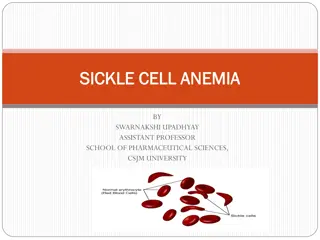
Anemia RBC loss without RBC Destruction Decreased RBC Production Increased RBC Destruction over Production (Hemolytic Anemias) Hemorrhage Due to trauma Due to disorders: e.g.cancer, ulcers Intrinsic Abnormalities Hereditary Spherocytosis Thalassemia Sickle Cell Anemia G6PD deficiency Hexokinase deficiency Neoplasia Leukemia Myelofibrosis Menstrual flow Pernicious anemia Gynecological disorders Iron Deficiency anemia Extrinsic Abnormalities Infections Malaria Mycoplasma - Parasitism Hookworms Aplastic anemia Chloramphenicol administration Renal disease (lack of erythropoietin production)
Basic Evaluation of Anemia Review of blood count, blood smear and RBC indices (MCV, MCH, MCHC) MCV is the most accurate method of measuring red blood cells and most useful in classification of anaemia as: Reticulocyte index = reticulocyte count (%) x [observed haematocrit / normal haematocrit] ie normalized for hematocrit Reticulocyte index > 2% indicates excessive RBC destruction or loss Reticulocyte index < 2% indicates decreased production
Effects of Anemia on Body Functions Increased cardiac output, as well as increased pumping workload on the heart, because of decreased viscosity and vasodilation. Reduced oxygen-carrying capacity of blood and lack of O2 for ATP and heat production, manifested mostly during exertion. If severe, can lead to heart failure
Clinical Picture of Anemia Symptoms fatigue, cold intolerance and paleness oxygen-carrying capacity of blood is reduced - lack of O2 for ATP and heat production Signs Pallor: an abnormal loss of skin or mucous membrane color. Koilonychia: is when the nail curves upwards (becomes spoon-shaped) Angular stomatitis: deep cracks and splits form at the corners of the mouth Glossitis: is inflammation or infection of the tongue
Polycythemia Types: True or absolute Primary (polycythemia rubra vera): uncontrolled RBC production Secondary to hypoxia: high altitude, chronic respiratory or cardiac disease Relative Hemoconcentration: loss of body fluid in vomiting, diarrhea, sweating Complications of polycythemia: hyperviscosity of the blood
Objectives; Intended learning outcomes (ILOs) After reviewing the PowerPoint presentation and the associated learning resources, the student should be able to: Discuss regulation of erythropoiesis Describe the factors required for normal erythropoiesis and the role of erythropoietin in erythropoiesis Recognize clinical conditions associated with high level of erythropoietin in the blood Summarize the synthesis of Hemoglobin Recognize hemoglobin structure and its functions Describe metabolism of iron and vitamin B12 Define anemia Classify anemia and explain its assessment Describe the physiological consequences and clinical picture of anemia Define polycythemia Classify polycythemia Describe the physiological consequences of polycythemia


")