Understanding Hepatobiliary and Pancreatic Disorders with Dr. Nadia Hameed
Hepatobiliary and pancreatic disorders can lead to conditions like jaundice, caused by an accumulation of bilirubin. Jaundice can be classified as prehepatic, intrahepatic, or posthepatic depending on the underlying causes such as liver damage, hepatitis, cirrhosis, or bile duct obstructions. Liver cirrhosis is a condition that changes the normal structure of the liver lobules, leading to issues like destruction of liver tissue and abnormal nodule formation. Cirrhosis can be categorized based on causative agents like biliary cirrhosis. Proper diagnosis involving bilirubin tests is crucial to determine the underlying cause of jaundice.

Download Presentation

Please find below an Image/Link to download the presentation.
The content on the website is provided AS IS for your information and personal use only. It may not be sold, licensed, or shared on other websites without obtaining consent from the author. Download presentation by click this link. If you encounter any issues during the download, it is possible that the publisher has removed the file from their server.
E N D
Presentation Transcript
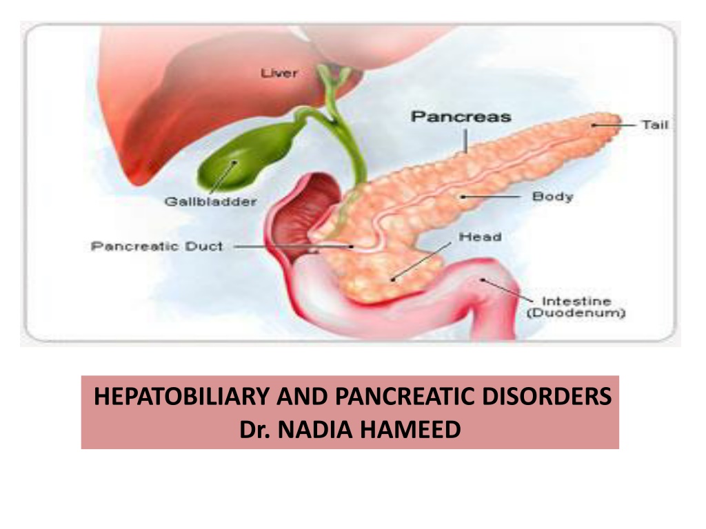
HEPATOBILIARY AND PANCREATIC DISORDERS Dr. NADIA HAMEED
Jundice (Icterus) Jaundice. Jaundice (i.e., icterus), which results from an abnormally high accumulation of bilirubin in the blood leading to yellowish discoloration to the skin, sclera and deep tissues. The hyperbilirubinemia may result from excessive destruction of red blood cells, impaired uptake of bilirubin by the liver cells, decreased conjugation of bilirubin, and obstruction of bile flow in the canaliculi of the hepatic lobules or in the intrahepatic or extrahepatic bile ducts. From an anatomic standpoint, jaundice can be categorized as prehepatic, intrahepatic, and posthepatic.
II// Intrahepatic Causes of Jaundice 1. Decreased bilirubin uptake by the liver I//Prehepatic (Excessive Red Blood Cell Destruction) 2. Decreased conjugation of bilirubin 1. Hemolytic blood transfusion reaction 3. Hepatocellular liver damage 2. Hereditary disorders of the red blood cell 4. Hepatitis 5. Cirrhosis 3. Sickle cell anemia 6. Cancer of the liver 4. Thalassemia 7. Drug-induced cholestasis 5. Spherocytosis III// Posthepatic (Obstruction of Bile Flow) 6. Acquired hemolytic disorders 1. Structural disorders of the bile duct 7. Hemolytic disease of the newborn 2. Congenital atresia of the extrahepatic bile ducts 8. Autoimmune hemolytic anemias 3. Bile duct obstruction caused by tumors or stone (cholelithiasis
Note: when the TSB lab test increased, conjugated and unconjugated bilirubin done: If the unconjugated bilirubin increased so the cause is prehepatic and hepatic. If the conjugated bilirubin is increased, so the cause is hepatic (after conjugation) and post hepatic.
Liver Cirrhosis Cirrhosis is a general term for a condition that destroys the normal architecture of the liver lobules. It structural features:- has the following important a. Destruction of liver parenchyma b. Separation of the lobules by fibrous tissues (by myofibroblast) activation of c. Formation of structurally abnormal nodules, and d. Abnormal vascular architecture
classifications of cirrhosis Cirrhosis is classified according to its causative agents and resultant pathologic configurations as :- a. Biliary cirrhosis b. post necrotic cirrhosis c. Alcoholic cirrhosis
a) Biliary cirrhosis - It is due to cholestasis which may be primary (idiopathic) or secondary to other conditions like bile duct obstruction or autoimmune diseases of the liver (autoimmune biliary cirrhosis) - The obstruction in one area of biliary passage results in bile stasis; that causes injury and scarring around the hepatocytes with evidence of fibrosis. b) Post necrotic cirrhosis - It follows massive liver necrosis and involves the destruction of lobules and even lobes of the liver. - It may occur after viral hepatitis, autoimmune hepatitis or after exposure to hepatotoxins such as certain drugs. c) Alcoholic cirrhosis - The most common cause of cirrhosis is excessive alcohol consumption - At least 75% of alcohol related deaths are attributed to cirrhosis.
Stages in developments of alcoholic cirrhosis 1. Stage of fatty change - Excessive accumulation of fat within liver cells causes liver enlargement - Alcohols replace fat as a fuel for liver metabolism and impair mitochondrial ability to oxidize fat. - Don t usually produce symptoms - It is reversible once the alcohol intake has been discontinued. 2. Stage of Alcoholic Hepatitis - It is an intermediate stage between fatty changes and cirrhosis - It is characterized by inflammation and necrosis of liver cells, thus is always serious and sometimes fatal. - The necrotic lesions are generally patchy but may involve entire lobe. - The stage is characterized by hepatic tenderness, paler, anorexia, nausea, jaundice, ascites and liver failure. Some patients may be asymptomatic. 3. Stage of cirrhosis Cirrhosis is the end result of liver injury caused by fatty liver and alcoholic hepatitis. The normal liver structure is replaced by bans of fibrous tissue with areas of regenerating cells. As the disease progress liver shrinks.
Clinical Manifestations of cirrhosis -The Manifestations Hepatomegally to hepatic failure. Early manifestations: - right upper quadrant pain - Sensation of fullness Late manifestation:- - The late manifestations are related to portal hypertension and liver cell failure ( Hepatocellular failure ) Portal Hypertension:- The fibrotic bands cause narrowing of the portal vein to cause portal hypertension. It s followed by back ward congestion of all tributaries of portal veins. Hepatocellular failure results in:- Decreased production of bile. Decreased plasma protein (Hypoalbuminemia) Decreased blood clotting factors. Accumulation of metabolic bi-products and toxins like bilirubin, ammonia and other substances in the circulation since the liver loses its detoxification capacity. This is one of the reasons for hepatic coma to occur. of cirrhosis are variable, ranging from asymptomatic
Liver Failure The most severe clinical consequence of liver disease is hepatic failure. It may result from sudden and massive hepatic destruction as in fulminant hepatitis, or it may be the result of progressive damage to the liver, such as occurs in alcoholic cirrhosis. Whatever the cause, 80% to 90% of hepatic functional capacity must be lost before hepatic failure occurs. The manifestations of liver failure reflect the various synthesis, storage, metabolic, and excretory functions of the liver. Fetor hepaticus refers to a characteristic musty, sweetish odor of the breath in the patient with advanced liver failure.
Cholelithiasis and Cholecystitis Two common disorders of the gallbladder system are cholelithiasis (i.e., gallstones) and inflammation of the gallbladder (cholecystitis) or common bile duct (cholangitis). Cholelithiasis Is the presence of one or more calculi (gallstones) in the gallbladder. In developed countries, about 10% of adults and 20% of people > 65 yr have gallstones. Gallstones tend to be asymptomatic. The most common symptom is biliary colic; gallstones do not cause dyspepsia or fatty food intolerance. More serious complications include cholecystitis; biliary tract obstruction, sometimes with infection (cholangitis); and gallstone pancreatitis. Diagnosis is usually by ultrasonography.
Three factors contribute to the formation of gallstones: (1) abnormalities in the composition of bile, (2) stasis of bile, as in pregnancy and parentral nutrition, dehydration(3) inflammation of the gallbladder. Three risk factors for gallstones include (1)female sex, (2)obesity, (3) family history. Most disorders of the biliary tract result from gallstones.
Pathophysiology of Cholelithiasis Biliary sludge is often a precursor of gallstones. It consists of Ca bilirubinate (a polymer of bilirubin), cholesterol microcrystals, and mucin. Sludge develops during gallbladder stasis, as occurs during pregnancy or use of TPN( total parenteral nutrition) . Most sludge is asymptomatic and disappears when the primary condition resolves. Alternatively, sludge can evolve into gallstones or migrate into the biliary tract, obstructing the ducts and leading to biliary colic, cholangitis, or pancreatitis. . Approximately 75% of gallstones are composed primarily of cholesterol; the other 25% are black or brown pigment stones consisting of calcium salts with bilirubin. many stones have a mixed composition.
Acute and Chronic Cholecystitis The term cholecystitis refers to inflammation of the gallbladder. Both acute and chronic cholecystitis are associated with cholelithiasis. Acute cholecystitis may be superimposed on chronic cholecystitis. Acute cholecystitis: acute inflammation of the gall bladder wall along with mucosal swelling and ischemia resulting from venous congestion and lymphatic stasis. The gallbladder usually is markedly distended and Bacterial infections may arise secondary to the ischemia and chemical irritation.
We have two types I// acute calculous cholecystitis: Referred to acute inflammation of the gall bladder that contain stones and is precipitated by obstruction of gallbladder neck or cystic duct. It is the most common complication of gallstones and the most common reason for emergency cholecystectomy. II// acute a calculous cholecystitis: Referred to acute inflammation of the gallbladder without stones. Occurs in 10% only and the patients are usually seriously ill eg after sever trauma, major surgery, burn and septicemia. In these cases dehydration, bile stasis and sludging occur.
pain becomes more pronounced in the right upper quadrant. The right subcostal region is tender (murphy sign) . Approximately 75% of patients have vomiting, and approximately 25% have jaundice. Fever high white blood cell by complete blood count. Total serum bilirubinTSB, aminotransferase, and alkaline phosphatase levels usually are elevated.
Chronic cholecystitis: Chronic cholecystitis results from repeated episodes of acute cholecystitis but in most instances it develops de novo. Chronic cholecystitis is almost always associated with gallstones but these do not seem to have direct role in the initiation of chronic inflammation. It is believed that supersaturation of bile predispose to both chronic inflammation and stones formation.
Acute pancreatitis represents an inflammation of the pancreasthat ranges from a mild self-limited disease, consisting of inflammation and interstitial edema, to an acute hemorrhagic pancreatitis that is associated with massive necrosis of tissue. Acute hemorrhagic pancreatitis is a severe, life- threatening disorder associated with the escape of activated pancreatic enzymes into the pancreas and surrounding tissues. Causes: 1. 20% idiopathic 2. most cases result from gallstones (stones in the common duct) 3. alcohol abuse These are the three most common causes. 4. Non gallstone obstruction of pancreatic duct eg by tumor 5. Drugs: thiazides and frusemide diuretics 6. Trauma: by accident or during surgery (iatrogenic) 7. Others like metabolic disorders, ischemia and infections especially with mump
Pathogenesis: A. Autodigestion of pancreatic substances by pancreatic enzymes. Trypsin have central role because it activate other enzymes (lipases and elastases) and activate kinin and factor XII which intern activate complement and clotting systems. B. Pancreatic duct obstruction allowing accumulation of an enzyme rich interstitial fluid that causes tissue injury and compromising blood flow. C. The alcoholic pancreatitis occur due to direct toxic effect of alcohol on pancreatic cells and alcohol consumption lead to contraction of sphincter of Oddi.
Clinical features: The most common initial symptom is severe epigastric and abdominal pain that radiates to the back. Tachycardia, hypotension, cool and clammy skin, and fever often are evident. Signs of hypocalcemia may develop, probably as a result of the precipitation of serum calcium in the areas of fat necrosis. Shock may develop due to electrolyte disturbance, increased vascular permeability and loss of blood. Lab findings: Hypocalcemia occurs in approximately 25% of patients. Total serum amylase is the test used most frequently in the diagnosis of acute pancreatitis. Serum amylase levels increase within the first 24 hours after onset of symptoms and remain elevated for 48 to 72 hours. The serum lipase level also increases during the first 24 to 48 hours and remains elevated for5 to 14 days.
Chronic pancreatitis: Is characterized by long standing inflammation and fibrosis with destruction of the exocrine pancreas. In the late stage the islets also destructed and lost. At this point, signs of diabetes mellitus and the malabsorption syndrome (e.g., weight loss, fatty stools [steatorrhea]) become apparent. Causes: a) Chronic alcoholism (the most common cause) b) Long standing pancreatic duct obstruction (by pseudocyst,calculi, neoplasm) c) Tropical pancreatitis: seen in Africa and Asia and attributed to malnutrition d) Hereditary pancreatitis due to mutation in genes encoding trypsin inhibitors.


")


 Biliary cirrhosis")