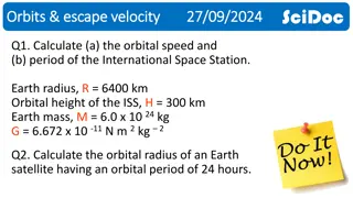
Understanding the Orbital Region: An Overview
The orbital region encompasses the orbits, eyelids, ciliary and tarsal glands, and anatomical borders. It consists of bony cavities protecting the eyeballs, eyelids that shield the eyes, and intricate structures like the lacrimal apparatus and muscles. Understanding the components and anatomical relations of the orbital region is crucial for comprehending eye anatomy and functionality. Learn more about this intricate area through detailed images and descriptions provided in this informative content.

Download Presentation

Please find below an Image/Link to download the presentation.
The content on the website is provided AS IS for your information and personal use only. It may not be sold, licensed, or shared on other websites without obtaining consent from the author. Download presentation by click this link. If you encounter any issues during the download, it is possible that the publisher has removed the file from their server.
E N D
Presentation Transcript
The orbital region Dr. Halah Abbass Lecture 2
The orbits The orbits are a pair of bony cavities that contain the eyeballs; their associated muscles, nerves, vessels, and fat; and most of the lacrimal apparatus. The orbital opening is guarded by two thin, movable folds, the eyelids.
Eyelids The eyelids protect the eye from injury and excessive light by their closure . The upper eyelid is larger and more mobile than the lower, and they meet each other at the medial and lateral angles. The palpebral fissure is the elliptical opening between the eyelids and is the entrance into the conjunctival sac. The superficial surface of the eyelids is covered by skin, and the deep surface is covered by a mucous membrane called the conjunctiva.
The ciliary glands (glands of Moll) are modified sweat glands that open separately between adjacent lashes. The tarsal glands (Meibomian glands) are long, modified sebaceous glands that pour their oily secretion onto the margin of the lid Near the medial angle of the eye a small elevation, the papilla lacrimalis, is present. On the summit of the papilla is a small hole, the punctum lacrimale, which leads into the canaliculus lacrimalis (Figs. 1 & 2). The punctum and canaliculus carry tears down into the nose.
Borders and Anatomical Relations Roof (superior wall) Formed by the frontal bone and the lesser wing of the sphenoid. The frontal bone separates the orbit from the anterior cranial fossa. Floor (inferior wall) Formed by the maxilla, palatine and zygomatic bones. The maxilla separates the orbit from the underlying maxillary sinus. Medial wall Formed by the ethmoid, maxilla, lacrimal and sphenoid bones. The ethmoid bone separates the orbit from the ethmoid sinus. Lateral wall Formed by the zygomatic bone and greater wing of the sphenoid. Apex Located at the opening to the optic canal, the optic foramen. Base Opens out into the face, and is bounded by the eyelids. It is also known as the orbital rim.
Movements of the Eyelids The eyelids are closed by the contraction of the orbicularis oculi and the relaxation of the levator palpebrae superioris muscles. The eye is opened by the levator palpebrae superioris raising the upper lid. On looking upward, the levator palpebrae superioris contracts, and the upper lid moves with the eyeball. On looking downward, both lids move, the upper lid continues to cover the upper part of the cornea, and the lower lid is pulled downward slightly by the conjunctiva, which is attached to the sclera and the lower lid.
Lacrimal Apparatus Lacrimal Gland consists of a large orbital part and a small palpebral part, which are continuous with each other around the lateral edge of the aponeurosis of the levator palpebrae superioris. It is situated above the eyeball and opens by 12 ducts into the lateral part of the superior fornix of the conjunctiva. The parasympathetic secretomotor nerve supply is derived from the lacrimal nucleus of the facial nerve. The sympathetic postganglionic nerve supply is from the internal carotid plexus and travels in the deep petrosal nerve, the nerve of the pterygoid canal, the maxillary nerve,the zygomatic nerve, the zygomaticotemporal nerve, and finally the lacrimal nerve. Lacrimal Ducts The tears circulate across the cornea and enter the canaliculi lacrimales through the puncta lacrimalis. The canaliculi lacrimales open into the lacrimal sac . The nasolacrimal duct emerges from the lower end of the lacrimal sac into the inferior meatus of the nose. The opening is guarded by a fold of mucous membrane known as the lacrimal fold.
Openings into the Orbital Cavity Supraorbital notch (Foramen): It is situated on the superior orbital margin and transmits the supraorbital nerve and blood vessels. Infraorbital groove and canal: Situated on the floor of the orbit in the orbital plate of the maxilla they transmit the infraorbital nerve and blood vessels. Nasolacrimal canal: Located anteriorly on the medial wall; it communicates with the inferior meatus of the nose It transmits the nasolacrimal duct. Superior orbital fissure: Located posteriorly between the greater and lesser wings of the sphenoid .It transmits the lacrimal nerve, the frontal nerve, the trochlear nerve, the oculomotor nerve (upper and 5 lower divisions), the abducent nerve, the nasociliary nerve, and the superior ophthalmic vein. Inferior orbital fissure: Located posteriorly between the maxilla and the greater wing of the sphenoid it communicates with the pterygopalatine fossa. It transmits the maxillary nerve and its zygomatic branch, the inferior ophthalmic vein, and sympathetic nerves. Optic canal: Located posteriorly in the lesser wing of the sphenoid .it communicates with the middle cranial fossa. It transmits the optic nerve and the ophthalmic artery.


 are modified sweat")