Understanding Swine Erysipelas: Causes, Symptoms, and Prevention
Swine Erysipelas, caused by Erysipelothrix rhusiopathiae, is a significant disease in pigs, characterized by cutaneous erythema, diamond-shaped lesions, septicemia, and arthritis. The infection is primarily transmitted through contaminated feed, water, or skin abrasions. Clinical findings include sudden deaths, febrile episodes, inappetence, painful joints, and distinctive skin discoloration. Prevention strategies involve good hygiene practices and vaccination programs.
Download Presentation

Please find below an Image/Link to download the presentation.
The content on the website is provided AS IS for your information and personal use only. It may not be sold, licensed, or shared on other websites without obtaining consent from the author. Download presentation by click this link. If you encounter any issues during the download, it is possible that the publisher has removed the file from their server.
E N D
Presentation Transcript
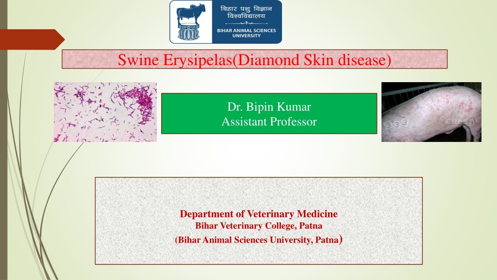
Swine Erysipelas(Diamond Skin disease) Dr. Bipin Kumar Assistant Professor Department of Veterinary Medicine Bihar Veterinary College, Patna (Bihar Animal Sciences University, Patna)
Swine Erysipelas(Diamond skin disease) Erysipelosis is caused primarily by Erysipelothrix rhusiopathiae, a bacteria carried by up to 50% of pigs. Clinically characterised by cutaneous erythema, including characteristic diamond- shaped lesions, septicemia, arthritis, and endocarditis.
Swine Erysipelas(Diamond skin disease) Erysipelosis is caused primarily by Erysipelothrix rhusiopathiae, a bacteria carried by up to 50% of pigs. Clinically characterised by cutaneous erythema, including characteristic diamond- shaped lesions, septicemia, arthritis, and endocarditis.
Swine Erysipelas(Diamond skin disease) Erysipelosis is caused primarily by Erysipelothrix rhusiopathiae, a bacteria carried by up to 50% of pigs. Clinically characterised by cutaneous erythema, including characteristic diamond- shaped lesions, septicemia, arthritis, and endocarditis.
Etiology and Pathogenesis Caused by E. rhusiopathiae in pigs. It is known as erysipelas and is one of the oldest recognized diseases of growing and adult swine. Up to 50% of pigs in intensive swine production areas are considered to be colonized with E rhusiopathiae. The organism commonly resides in the tonsillar tissue. These typical healthy carriers can shed the organism in their feces or oronasal secretions. Infection is by ingestion of contaminated feed, water, or feces and through skin abrasions. On farms where the organism is endemic, pigs are exposed naturally to E rhusiopathiae when they are young. Maternal derived antibodies provide passive immunity and suppress clinical
Clinical Findings Disease outbreaks may be acute or chronic, and clinically inapparent infections also occur. Acute outbreaks are characterized by; - Sudden and unexpected deaths - Febrile episodes,Inappetence,Painful joints - Skin lesions that vary from generalized cyanosis to the often-described diamond skin (rhomboid urticaria) lesions. - Skin discoloration may vary from widespread erythema and purplish discoloration of the ears, snout, and abdomen, to diamond-shaped skin lesions almost anywhere on the body, but particularly on the lateral and dorsal regions. Chronic form tends to follow acute outbreaks and is characterized by; - Enlarged joints and lameness.
Lesions At necropsy, acutely infected pigs may exhibit skin lesions, enlarged and congested lymph nodes, edematous and congested lungs, splenomegaly, and hepatomegaly. Petechial hemorrhages may be seen on the kidneys and heart. In chronic erysipelas, valvular endocarditis is seen as proliferative, granular growths on the heart valves. Arthritis may involve joints of one or more legs, and the intervertebral articulations. Proliferation and erosion of articular cartilage may result in fibrosis and ankylosis of the joint.
Diagnosis Clinical signs and/or gross lesions Response to antimicrobial therapy Demonstration of the bacterium or DNA in tissues (bacterial culture and/or molecular tests) Rhomboid urticaria or diamond skin lesions are almost diagnostic when present; however, similar lesions can also be seen with classical swine fever virus infection, Actinobacillus suis septicemia, or the porcine dermatitis and nephropathy syndrome.
Treatment Preventive treatment through vaccination should be emphasized. Early treatment with appropriate antibiotics, particularly penicillin, generally leads to recovery E rhusiopathiae is sensitive to penicillin. On an economic basis, penicillin is the best choice for antibiotic therapy, but ampicillin and ceftiofur also yield satisfactory results in acute cases. When injecting large numbers of affected pigs is impractical, tetracyclines delivered in the feed or water may be useful. Fever associated with acute infections can be managed by administration of NSAIDs such as flunixin meglumine or by delivery of aspirin in the water. Erysipelas antiserum is described as an effective adjunct to antibiotic therapy in treating acute outbreaks but is not commonly available. Treatment of chronic infections is usually ineffective and not cost effective.
Prevention Vaccination against E rhusiopathiae is very effective Injectable bacterins and attenuated, live vaccines delivered via the water are available and provide extended duration of immunity. Susceptible pigs may be vaccinated before weaning, at weaning, or several weeks after weaning. Male and female swine selected for addition to the breeding herd should be vaccinated with a booster 3 5 weeks later. Vaccination failures may occur in some herds. The use of live vaccines may lead to clinical disease, particularly chronic erysipelas. In addition to vaccination, attention to sanitation and hygiene and elimination of pigs with clinical signs suggestive of erysipelas infection represent other viable methods that may help control the disease on swine farms.
